Welcome to the Amira-Avizo Software Use Case Gallery
Below you will find a collection of use cases of our 3D data visualization and analysis software. These use cases include scientific publications, articles, papers, posters, presentations or even videos that show how Amira-Avizo Software is used to address various scientific and industrial research topics.
Use the Domain selector to filter by main application area, and use the Search box to enter keywords related to specific topics you are interested in.
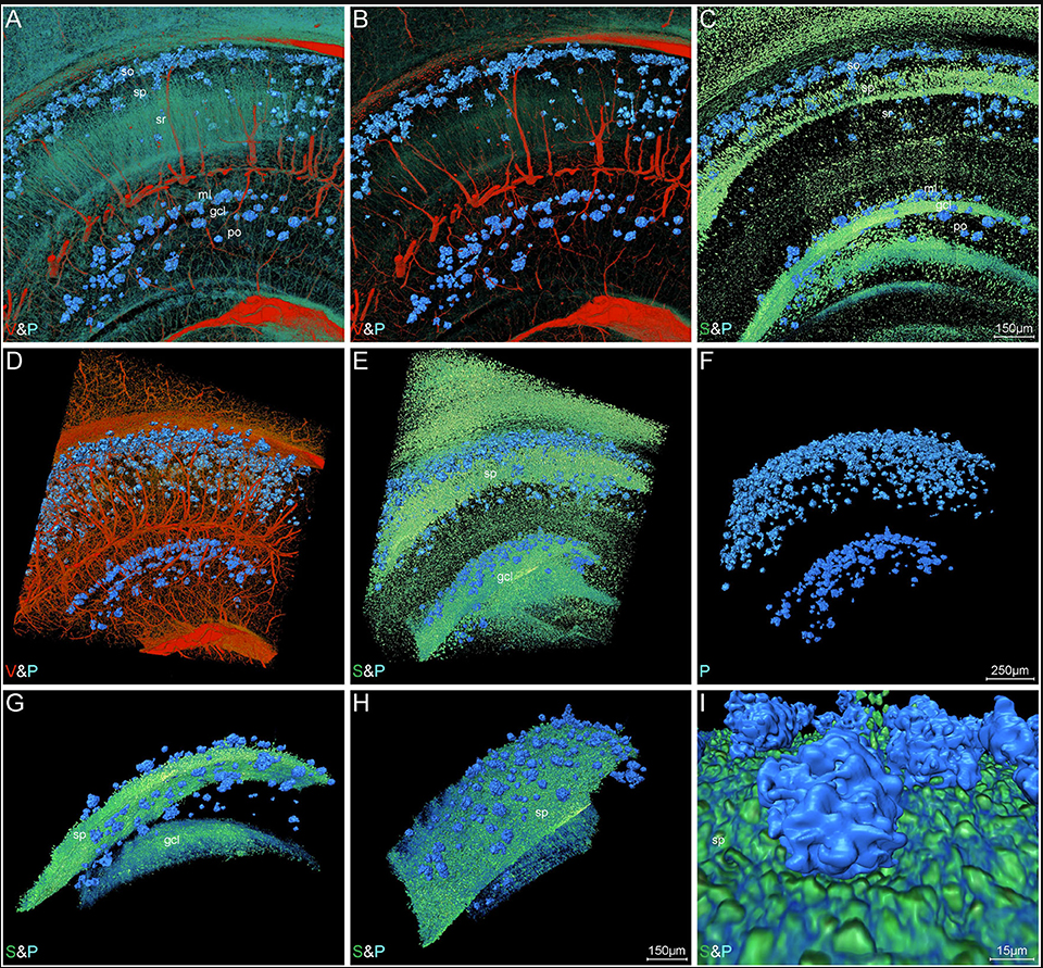
High-Resolution Digital Panorama of Multiple Structures in Whole Brain of Alzheimer's Disease Mice
Our study placed emphasis on solving problems in processing high-throughput bright field images and made attempt in developing a method for the extraction and reconstruction of multiple structures. This will facilitate a better understanding of the cerebral anatomical features under the pathological state of AD and shows extensive application prospect in drug efficacy assessment from brain-wide level.
Simultaneously visualizing Amyloid-β (Aβ) plaque with its surrounding brain structu... Read more
Xianzhen Yin, Xiaochuan Zhang, Jingjing Zhang, Weicheng Yang, Xian Sun, Haiyan Zhang, Zhaobing Gao, Hualiang Jiang
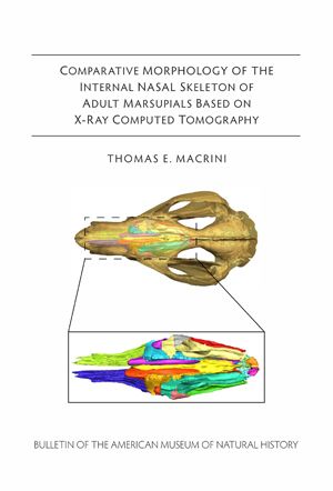
The internal skeleton of the nasal cavity is sporadically and often incompletely described for many marsupial species and mammals in general. Here, I provide an anatomical survey of the ethmoid in the skulls of adult marsupials based on examination of computed tomography (CT) imagery of 29 taxa representing all the major extant groups of marsupials. (…) I compared ecological data with the number of ecto- and endoturbinals as a preliminary test to determine whether some of the interspec... Read more
Thomas E. Macrini
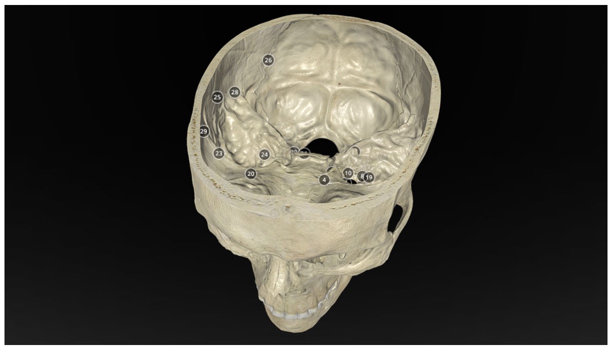
Operative Anatomy of the Human Skull: A Virtual Reality Expedition
Benjamin K Hendricks, MD Akash J Patel, MD Jerome Hartman Mark F Seifert, PhD Aaron Cohen-Gadol, MD, MSc, MBA
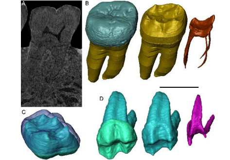
Exploring hominin and non-hominin primate dental fossil remains with neutron microtomography
Fossil dental remains are an archive of unique information for paleobiological studies. Computed microtomography based on Xray microfocus sources (X-µCT) and Synchrotron Radiation (SR-µCT) allow subtle quantification at the micron and sub-micron scale of the meso- and microstructural signature imprinted in the mineralized tissues, such as enamel and dentine, through highresolution “virtual histology”. Nonetheless, depending on the degree of alterations undergone during fossiliza... Read more
Clément Zanolli, Laboratory AMIS, UMR 5288, University of Toulouse III - Paul Sabatier, France, and al.
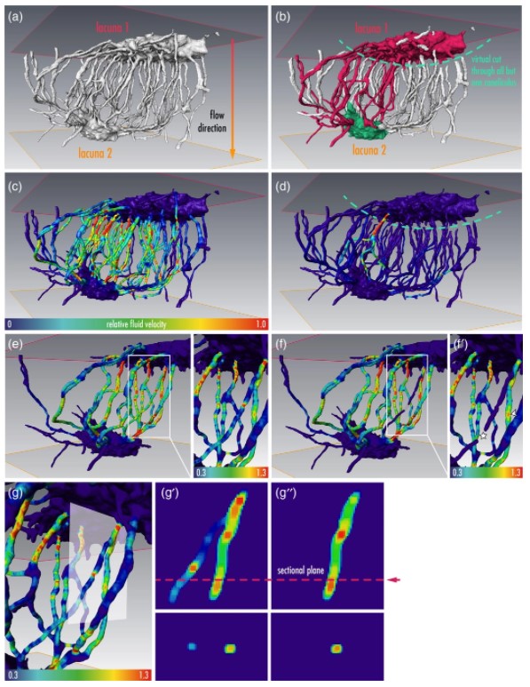
Osteocytes are the most frequent bone cells connected with each other through cell processes within tiny tubular-shaped canaliculi. The so-called osteocyte lacunar-canalicular network (LCN) plays a crucial role in bone remodeling and mineral homeostasis. Given the critical nature of these functions, it is herein hypothesized that the LCN must be structurally “overengineered” to provide network resilience.
This hypothesis is tested by characterizing canalicular networks in human bon... Read more
Emely Bortel, Liam M Grover, Neil Eisenstein, Christian Seim, Heikki Suhonen, Alexandra Pacureanu, Peter Westenberger, Kay Raum, Max Langer, Francoise Peyrin, Owen Addison, Bernhard Hesse
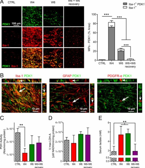
Proinflammatory mononuclear phagocytes (MPs) play a crucial role in the progression of multiple sclerosis (MS) and other neurodegenerative diseases. Despite advances in neuroimaging, there are currently limited available methods enabling noninvasive detection of MPs in vivo. Interestingly, upon activation and subsequent differentiation toward a proinflammatory phenotype MPs undergo metabolic reprogramming that results in increased glycolysis and production of lactate. Hyperpolarized (HP)
Caroline Guglielmetti, Chloé Najac, Alessandro Didonna, Annemie Van der Linden, Sabrina M. Ronen, and Myriam M. Chaumeil
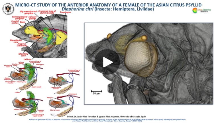
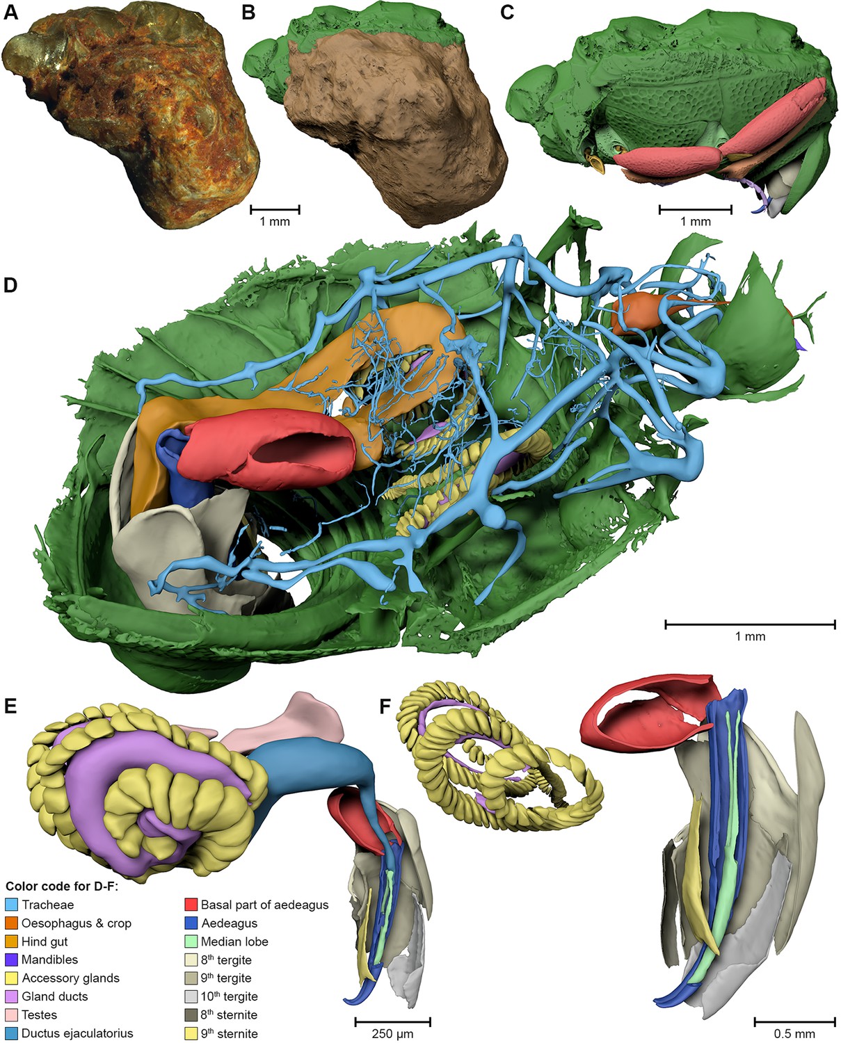
The Asian citrus psyllid (ACP), Diaphorina citri, is a harmful pest of citrus trees that transmits Candidatus Liberibacter spp. which causes Huanglongbing (HLB) (citrus greening disease); this is considered to be the most serious bacterial disease of citrus plants.
Here we detail an anatomical study of the external and internal anatomy (excluding the reproductive system) using micro-computed tomography (micro-CT). This is the first complete 3D micro-CT reconstruction o... Read more
Javier Alba-Tercedor, Wayne B. Hunter & Ignacio Alba-Alejandre
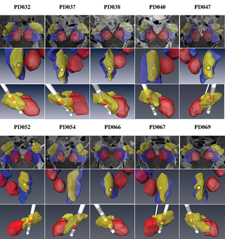
Patient-specific anatomical model for deep brain stimulation based on 7 Tesla MRI
Deep brain stimulation (DBS) requires accurate localization of the anatomical target structure, and the precise placement of the DBS electrode within it. Ultra-high field 7 Tesla (T) MR images can be utilized to create patient-specific anatomical 3D models of the subthalamic nuclei (STN) to enhance pre-surgical DBS targeting as well as post-surgical visualization of the DBS lead position and orientation. We validated the accuracy of the 7T imaging-based patient-specific model of the STN and m... Read more
Yuval Duchin, Reuben R. Shamir, Remi Patriat, Jinyoung Kim, Jerrold L. Vitek, Guillermo Sapiro, Noam Harel
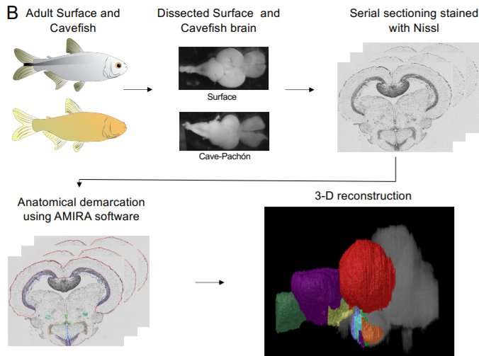
A shift in environmental conditions impacts the evolution of complex developmental and behavioral traits. The Mexican cavefish, Astyanax mexicanus, is a powerful model for examining the evolution of development, physiology, and behavior because multiple cavefish populations can be compared to an extant and ancestral-like surface population of the same species. Many behaviors have diverged in cave populations of A. mexicanus, and previous studies have shown that cavefish ha... Read more
Cody Loomis, View ORCID ProfileRobert Peuß, James Jaggard, Yongfu Wang, Sean McKinney, Stephen Raftopoulos, Austin Raftopoulos, Daniel Whu, Matthew Green, Suzanne E. McGaugh, Nicolas Rohner, Alex C. Keene, Erik R. Duboue

Each pulmonary segment is an anatomical and functional unit. However, it is fundamentally difficult to precisely distinguish every pulmonary segment using the conventional pulmonary intersegmental planes from computed tomography images. Building arteriopulmonary segments is likely to be an effective way to identify pulmonary segments.
The three-dimensional reconstructed images showed the branches of the pulmonary artery ramified up to their eighth order covering the entire lung as well... Read more
Huijie Gao, Chao Liu
Progressive transformation of the otic placode into the functional inner ear during gestational development in humans leads to the acquisition of hearing perception via the cochlea and balance and spatial orientation via the vestibular organ.
Using a correlative approach involving micro-computerized tomography (micro-CT), transmission electron microscopy and histological techniques we were able to examine both the morphological and cellular changes associated with human inner ear devel... Read more
Lejo Johnson Chacko, David Wertjanz, Consolato Sergi, Jozsef Dudas, Natalie Fischer, Theresa Eberharter, Romed Hoermann, Rudolf Glueckert, Helga Fritsch, Helge Rask-Andersen, Anneliese Schrott-Fischer & Stephan Handschuh
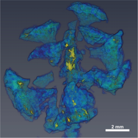
3D computational anatomy of the scaphoid and its waist for use in fracture treatment
A detailed understanding of scaphoid anatomy helps anatomic fracture reduction and optimal screw position. Therefore, we analyzed the size and shape variations of the cartilage and osseous surface, the distribution of volumetric bone mineral density (vBMD), and if the vBMD values differ between a peripheral and a central screw pathway?
Forty-three fresh frozen hand specimens (17 females, 26 males) were analysed with high-resolution peripheral quantitative computed tomography (HR-pQCT) ... Read more
Marc-Daniel Ahrend, Teun Teunis, Hansrudi Noser, Florian Schmidutz, Geoff Richards, Boyko Gueorguiev & Lukas Kamer
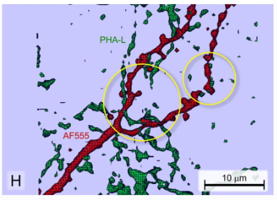
Neuroanatomical tract-tracing techniques that did go viral
Neuroanatomical tracing methods remain fundamental for elucidating the complexity of brain circuits. During the past decades, the technical arsenal at our disposal has been greatly enriched, with a steady supply of fresh arrivals. This paper provides a landscape view of classical and modern tools for tract-tracing purposes. Focus is placed on methods that have gone viral, i.e., became most widespread used and fully reliable.
Read more
Jose L. Lanciego; Floris G. Wouterlood
Juvenile Ovine Ex Vivo Larynges: Phonatory, Histologic, and Micro CT Based Anatomic Analyses
It is well known that the phonatory process changes during the life span. However, detailed investigations on potential factors concerned are rare. To deal with this issue, we performed extended biomechanical, macro anatomical, and histological analyses of the contributing laryngeal structures in ex vivo juvenile sheep models. Altogether twelve juvenile sheep larynges were analyzed within the phonatory experiments. Three different elongation levels and 16 different flow levels were applied to... Read more
Michael Döllinger, Olaf Wendler, Claus Gerstenberger, Tanja Grossmann, Marion Semmler, Hossein Sadeghi, and Markus Gugatschka
Computed tomography is an increasingly popular technique for the non-destructive study of fossils. Whilst the science of X-ray computed tomography (CT) has greatly matured since its first fossil applications in the early 1980s, the applications and limitations of neutron tomography (NT) remain relatively unexplored in palaeontology. These highest resolution neutron tomographic scans in palaeontology to date were conducted on a specimen of Austrosequoia novae-zeelandiae (Ettingshausen) Mays an... Read more
Chris Mays, Joseph J. Bevitt, and Jeffrey D. Stilwell
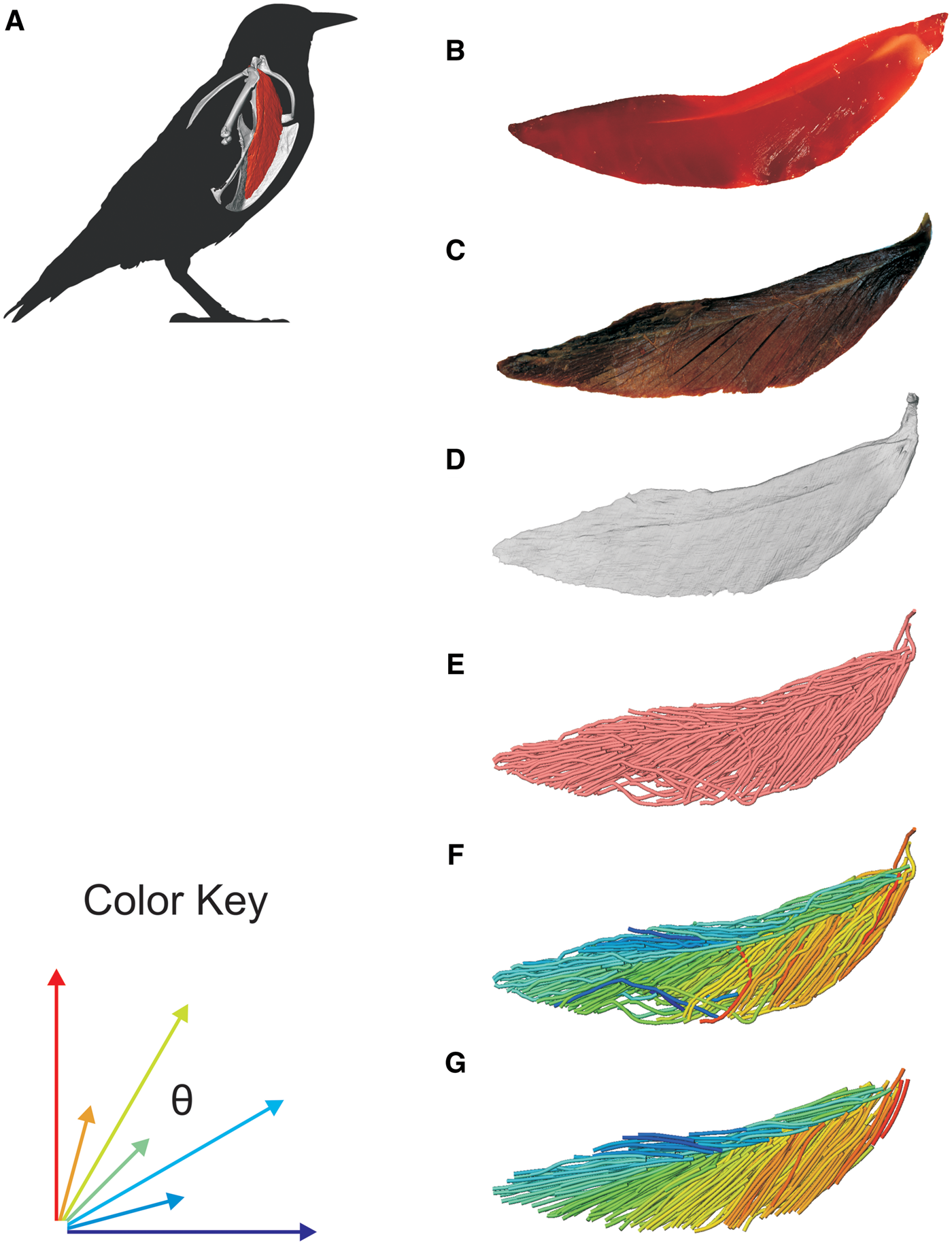
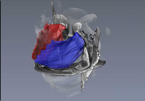
3D Muscle Architecture of the Pectoral Muscles of European Starling (Sturnus vulgaris)
Avian flight is achieved through a number of modifications to the body, including the pectoral girdle (…). Muscle architecture is a critical variable in determining the biomechanical function of the vertebrate musculoskeletal system; however, accurate three-dimensional (3D) understanding of muscle architecture has been historically difficult to acquire. Here, we present a musculoskeletal model of a European starling (Sturnus vulgaris) pectoral girdle generated from iodine contr... Read more
S.P. Sullivan, F.R. McGechie, K.M. Middleton, C.M. Holliday
External and internal morphological characters of extant and fossil organisms are crucial to establishing their systematic position, ecological role and evolutionary trends. (…) We found well-preserved three-dimensional anatomy in mineralized arthropods from Paleogene fissure fillings and demonstrate the value of these fossils by utilizing digitally reconstructed anatomical structure of a hister beetle. The new anatomical data facilitate a refinement of the species diagnosis and allowed... Read more
Achim H Schwermann, Tomy dos Santos Rolo, Michael S Caterino, Gunter Bechly, Heiko Schmied, Tilo Baumbach, Thomas van de Kamp
University of York student wins Anatomical Society Best Image Prize using Amira-Avizo Software
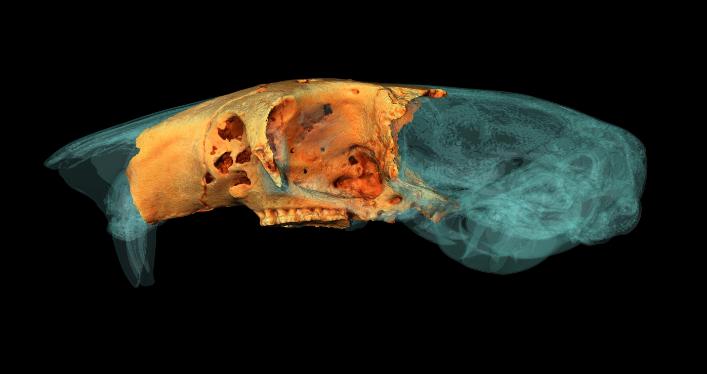
PhD student Jesse Hennekam wins for his reconstruction of the skull of a giant dormouse.
A York PhD student has won a prestigious award for his work reconstructing the skull of a giant rodent.
Jesse Hennekam, from the Centre for Anatomical and Human Sciences at the Hull York Medical School, created a digital reconstruction of the skull of a gigantic dormouse (Leithia melitensis), which roamed on the island of Sicily during the Pleistocene, roughly 2 million years ago... Read more
Jesse Hennekam
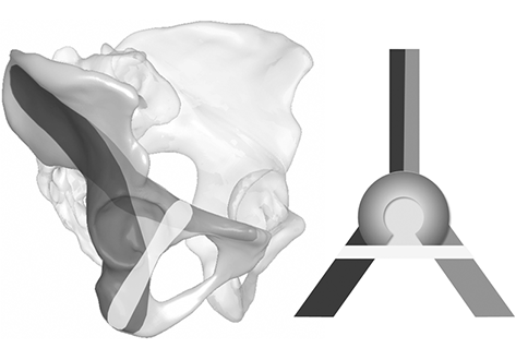
Acetabular fracture surgery is directed toward anatomical reduction and stable fixation to allow for the early functional rehabilitation of an injured hip joint. Recent biomechanical investigations have shown the superiority of using an additional screw in the infraacetabular (IA) region, thereby transfixing the separated columns to strengthen the construct by closing the periacetabular fixation frame. However, the inter-individual existence and variance concerning secure IA screw corridors a... Read more
Stephan Arlt, Hansrudi Noser, Andreas Wienke, Florian Radetzki, Gunther Olaf Hofmann, Thomas Mendel
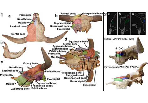
The Niata was a cattle variety from South America that figured prominently in writings on evolution by Charles Darwin. Its shortened head and other aspects of its unusual morphology have been subject of unsettled discussions since Darwin’s time. Here, we examine the anatomy, cranial shape, skull biomechanics, and population genetics of the Niata. Our results show that the Niata was a viable variety of cattle and exhibited anatomical differences to known chondrodysplastic forms. In cranial s... Read more
Kristof Veitschegger, Laura A. B. Wilson, Beatrice Nussberger, Glauco Camenisch, Lukas F. Keller, Stephen Wroe, Marcelo R. Sánchez-Villagra
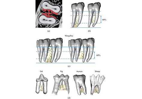
Mechanical adaptation of trabecular bone morphology in the mammalian mandible
Alveolar bone, together with the underlying trabecular bone, fulfils an important role in providing structural support against masticatory forces. Diseases such as osteoporosis or periodontitis cause alveolar bone resorption which weakens this structural support and is a major cause of tooth loss. However, the functional relationship between alveolar bone remodelling within the molar region and masticatory forces is not well understood. This study investigated this relationship by comparing m... Read more
Peter J. Watson, Laura C. Fitton, Carlo Meloro, Michael J. Fagan, Flora Gröning